Question
In: Physics
With respect to magnetic resonance imaging, describe the role of the following in producing an image:...
Solutions
Expert Solution
a) The Role of the Fixed Magnetic Field
The magnet is the most important and
biggest part of the MRI device. It is this
magnet that allows the MRI machine to produce high quality
images.MRI uses magnetic fields and radio waves to measures how
much water is in different tissues of the body, maps the location
of the water and then uses this information to generate a detailed
image. The images are so detailed because our bodies are made up of
around 65% water, so we have lots of signal to measure
There is a horizontal tube that runs through the magnet and is
called a bore. The magnet
is extremely powerful and its strength is measured in either
‟teslaˮ or ‟gaussˮ
(1 tesla = 10 000 gauss). Most MRI magnets use a magnetic field of
0.5 to 2.0
tesla, when the Earth’s magnetic field is only 0.5 gauss. The
magnetic field is
produced by passing current through multiple coils that are inside
the magnet,
resulting in a state of superconductivity, which produces a lot of
energy by
reducing the resistance in the wires to zero.
b) Role of the magnetic field gradients
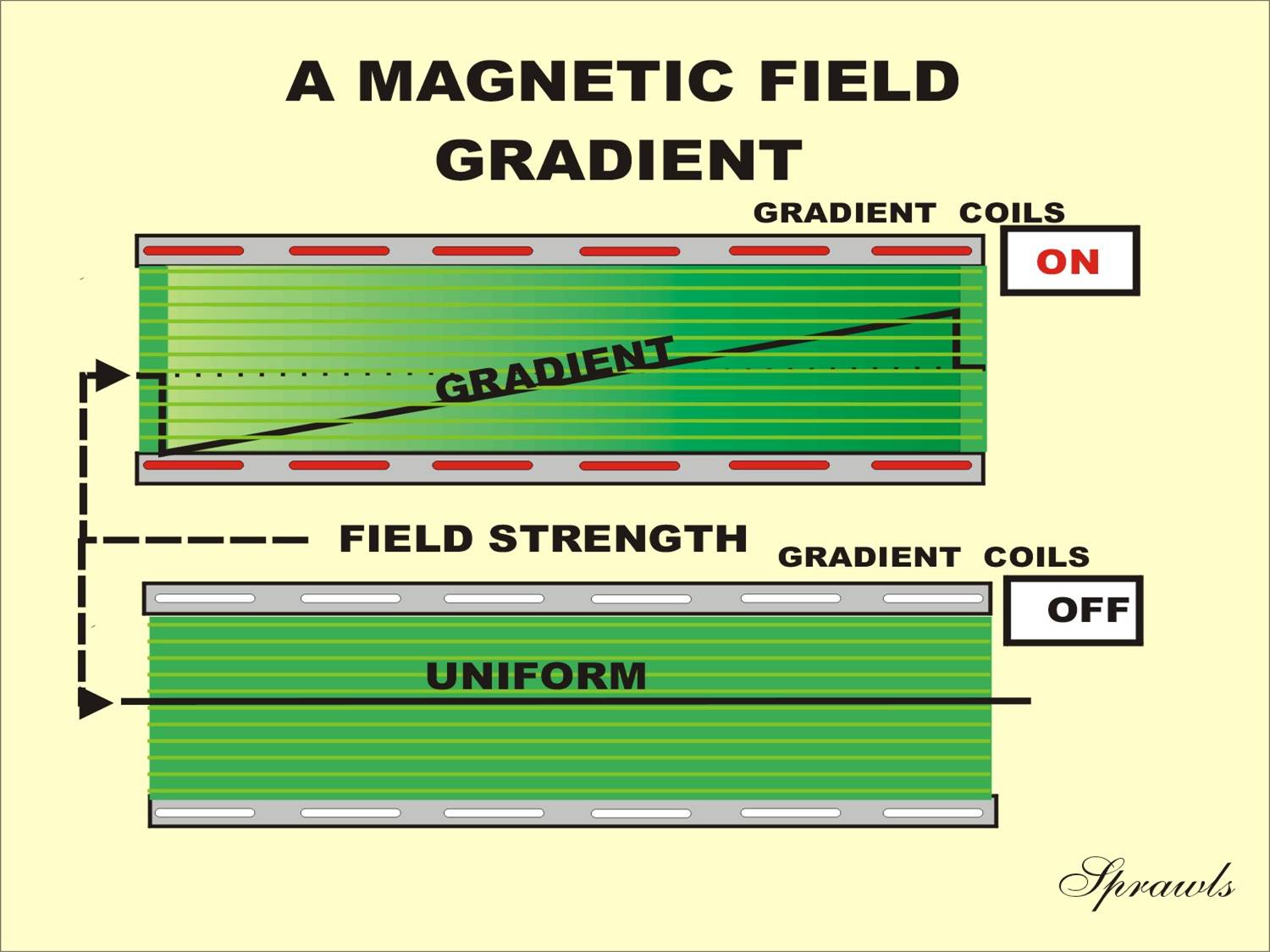
When the MRI system is in a resting state and not actually producing an image, the magnetic field is quite uniform or homogeneous over the region of the patient’s body. However, during the imaging process the field must be distorted with gradients. A gradient is just a change in field strength from one point to another in the patient’s body. The gradients are produced by a set of gradient coils, which are contained within the magnet assembly. During an imaging procedure the gradients are turned on and off many times. This action produces the sound or noise that comes from the magnet.
The effect of a gradient is illustrated in Figure 2-4. When a magnet is in a “resting state,” it produces a magnetic field that is uniform or homogenous over most of the patient’s body. In this condition there are no gradients in the field. However, when a gradient coil is turned on by applying an electric current, a gradient or variation in field strength is produced in the magnetic field.
|
Figure 2-4. |
 |
c) Role of radiofrequency field
The radio frequency (RF) system provides the communications link with the patient’s body for the purpose of producing an image. All medical imaging modalities use some form of radiation (e.g., x-ray, gamma-ray, etc.) or energy (e.g., ultrasound) to transfer the image from the patient’s body.
The MRI process uses RF signals to transmit the image from the patient’s body. The RF energy used is a form of non-ionizing radiation. The RF pulses that are applied to the patient’s body are absorbed by the tissue and converted to heat. A small amount of the energy is emitted by the body as signals used to produce an image. Actually, the image itself is not formed within and transmitted from the body. The RF signals provide information (data) from which the image is reconstructed by the computer. However, the resulting image is a display of RF signal intensities produced by the different tissues.
RF Coils
The RF coils are located within the magnet assembly and relatively close to the patient’s body. These coils function as the antennae for both transmitting signals to and receiving signals from the tissue. There are different coil designs for different anatomical regions (shown in Figure 2-6). The three basic types are body, head, and surface coils. The factors leading to the selection of a specific coil will be considered in Chapter 10. In some applications the same coil is used for both transmitting and receiving; at other times, separate transmitting and receiving coils are used.
|
Figure 2-6. The three types of RF coils (body, head, and
surface) that are the antennae |
 |
Surface coils are used to receive signals from a relatively small anatomical region to produce better image quality than is possible with the body and head coils. Surface coils can be in the form of single coils or an array of several coils, each with its own receiver circuit operated in a phased array configuration. This configuration produces the high image quality obtained from small coils but with the added advantage of covering a larger anatomical region and faster imaging.
Transmitter
The RF transmitter generates the RF energy, which is applied to the coils and then transmitted to the patient’s body. The energy is generated as a series of discrete RF pulses. As we will see in Chapters 6, 7, and 8, the characteristics of an image are determined by the specific sequence of RF pulses.
The transmitter actually consists of several components, such as RF modulators and power amplifiers, but for our purposes here we will consider it as a unit that produces pulses of RF energy. The transmitters must be capable of producing relatively high power outputs on the order of several thousand watts. The actual RF power required is determined by the strength of the magnetic field. It is actually proportional to the square of the field strength. Therefore, a 1.5 T system might require about nine times more RF power applied to the patient than a 0.5 T system. One important component of the transmitter is a power monitoring circuit. That is a safety feature to prevent excessive power being applied to the patient’s body, as described in Chapter 15.
Receiver
A short time after a sequence of RF pulses is transmitted to the patient’s body, the resonating tissue will respond by returning an RF signal. These signals are picked up by the coils and processed by the receiver. The signals are converted into a digital form and transferred to the computer where they are temporarily stored.
RF Polarization
The RF system can operate either in a linear or a circularly polarized mode. In the circularly polarized mode, quadrature coils are used. Quadrature coils consist of two coils with a 90˚ separation. This produces both improved excitation efficiency by producing the same effect with half of the RF energy (heating) to the patient, and a better signal-to-noise ratio for the received signals.
RF Shielding
RF energy that might be in the environment could be picked up by the receiver and interfere with the production of high quality images. There are many sources of stray RF energy, such as fluorescent lights, electric motors, medical equipment, and radio communications devices. The area, or room, in which the patient’s body is located must be shielded against this interference.
An area can be shielded against external RF signals by surrounding it with an electrically conducted enclosure. Sheet metal and copper screen wire are quite effective for this purpose.
The principle of RF shielding is that RF signals cannot enter an electrically conductive enclosure. The thickness of the shielding is not a factor—even thin foil is a good shield. The important thing is that the room must be completely enclosed by the shielding material without any holes. The doors into imaging rooms are part of the shielding and should be closed during image acquisition.
c)
A flip angle of less than 90° only
partially converts the z-magnetization, leaving a fraction cos
 along the longitudinal direction. A flip angle of 90° converts all
the z-magnetization into xy-magnetization.
When the repetition time is shorter than T1, the use of a partial flip angle can lead to higher signal intensity. The maximum signal intensity is given by the Ernst angle. For spin echo pulse sequences using an odd number of 180° pulses, an effect similar to the use of a partial flip angle is obtained by using a flip angle greater than 90° to offset the inversion of the remaining longitudinal magnetization by the 180° pulse
d) MRI head coils / MRI brain coils are typically birdcage coils. Multi channel coils allow to speed up the scan time with parallel imaging particularly at 3T (coils with 8 to 16 channel / elements are common).
Related Solutions
what is the importance and features of magnetic resonance imaging
Explain Magnetic Resonance Imaging.What is the fundamental physics concepts behind Magnetic Resonance Imaging? How does it...
why would someone be curious about Nuclear Magnetic Resonance (NMR) and Magnetic Resonance Imaging (MRI)? and...
i) Explain the working principle of Magnetic resonance imaging ii) What is the main advantage to...
29C MRI Magnetic Resonance Imaging takes exquisite images of the brain and other parts of human...
You recently requested bids for a new MRI (magnetic resonance imaging) machine in for your radiology...
Magnetic Resonance Spectroscopy (MRS) has the potential to provide whole body molecular imaging without injection of...
3) The very first magnetic resonance imaging (MRI) devices employed multi-layer copper solenoids, like the one...
PHYS/BIOL 464 Medical Imaging Instrumentation MRI HW 7A 1.During the magnetic resonance relaxation process after a...
How does magnetic resonance image (MRI) allow us to see details in soft nonmagnetic tissue? Use/connect...
- On June 30, Sharper Corporation’s stockholders' equity section of its balance sheet appears as follows before...
- In this journal you are asked to take the role of a mayor or congressional representative...
- Answer correctly the below 25 multiple questions on Software Development Security. Please I will appreciate the...
- 1. The activation energy of a certain reaction is 41.5kJ/mol . At 20 ?C , the...
- Give TWO pieces of evidence that you've successfully made methyl salicylate. Remember when you cite TLC...
- Describe briefly the evolution of Craniata and Vertebrata.
- How many grams are in a 0.10 mol sample of ethyl alcohol?
 genius_generous answered 3 years ago